

Microplate readers, also known as microplate photometers, are pieces of equipment that are utilized in the process of identifying biological, chemical, or physical occurrences that take place in the material that is contained on microtiter plates. The pharmaceutical and biotechnology industries, in addition to academic institutions, make extensive use of them in research, the discovery of new drugs, the validation of bioassays, quality control, and manufacturing processes. Microplate readerWhen conducting biochemical assays in the laboratory, microplate readers are the instruments that are typically used. These readers are used to determine the total phenolic content, total flavonoid content, and total tannin content. The most common type of microplate format found in university research labs and clinical diagnostic labs is the 96-well (8 by 12-matrix) microplate. A volume of between 100 and 200 L is typically used for the reaction in each well. The basic idea behind the microplate readerMany people believe that a spectrophotometer with microplate reading capabilities is the same thing as a microplate reader. The only thing that differentiates the traditional spectrophotometer and the microplate reader is the presence of colorimetric agents in the microplate reader. Traditional spectrophotometers don't have this feature. The conventional spectrophotometer has a greater range of wavelengths to read than the multi-plate reader does.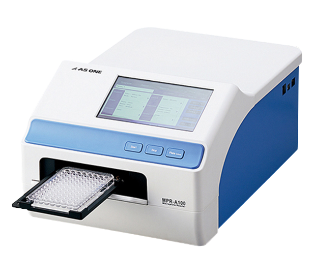
In addition, the results can be interpreted with the help of the microplate reader's diffracting gratings. This is especially significant in the Elisa approaches for the identification of the many different types of cancers. A wavelength range of 400–750 nm is found in the most sophisticated microplate readers. Nevertheless, it cannot be denied that some microplate readers perform analysis with the aid of UV light. Light with wavelengths ranging from 340 to 700 nm is utilized for the analysis. They make use of optical fibers in order to deliver light rays to the plate reader wells that contain biological samples. In order to conduct an analysis, these specimens or samples are loaded into the microplate reader and placed in one of its wells. The laser beams' diameters range from one millimeter to three millimeters as they travel through the biological samples. A system known as an ELISA plate reader is used in order to make a detection of the light rays that are produced as a result of the sample reactions. Amplification of the signal and determination of the absorbance rate of the reaction sample both take place during this step.
After that, the light beams are read by plate readers, which are part of the reading system, so that the data can be interpreted in a way that is both clear and precise. Additionally, Microplate reader enables the precise interpretation of test results based on a sample. Evaluations that need to be carried outIn order to use the microplate reader, one must first conduct a few tests. In many instances, the three functions of absorbance, fluorescence, and luminescence serve as the focal points of the most typical reading modes.1. Absorbance microplate readerA light source that emits light at a specific wavelength is used in this method of microplate detection. This light source is used to illuminate the biological response specimen. To select the desired wavelength, either a monochromator or an optical filter will do the trick here. A light ray detector is located on the opposite side of the microwell in the plate, and it is used to determine how much of the initial light passes through the biological sample. It is common practice to find a correlation between the amount of light that is transmitted and the concentration of the target molecule that is present within the wells. Additional conventional colorimetric analyses have been added to the wells of an ELISA microplate reader so that the samples can be examined in the most accurate manner possible.
In addition to this, its size has been drastically decreased so that microplate reader can perform quantitatively in the very best microplate reader while still maintaining a level of performance that is suitable for the requirements of biological research. Note: ABS = absorbance, LUM= luminescence, FL= fluorescence2. Fluorescence microplate readerIn the past two decades of biological research, fluorescence intensity has been a significant method in the microplate reading format. This format is used to analyze samples. The microplate absorbance detection method has a limited number of applications, whereas the fluorescence intensity detection method has a significantly wider range of potential uses. On the other hand, the equipment used for fluorescence detection is typically more expensive. The following are the components that make up the fluorescence intensity instrumentation found in this type of microplate reader:
The excitation system shines light of a specific wavelength onto the sample, which causes microplate reader to become illuminated
- The selection is typically done with a monochromator or an optical filter
- In addition to that, there is the lighting system
- As a result of the light rays passing through the specimen, a phenomenon known as fluorescence occurs
- The microplate reader's emission system is the optical system, which collects the light that has been emitted and separates it from the light that has been used to excite the plate
- This separation is achieved through the utilization of the monochromator system
- In addition, a light detector that is referred to as a photomultiplier tube (PMT) is utilized in order to measure the signal
- 3
- Choosing a microplate reader according to the total number of wells in the plate1
- Plate Reader with a Range of 6 to 96
This multimode plate reader has up to 96 wells that can be used for the storage of reaction samples. Because of this, detection is an easy process to carry out. It is possible to read all detections by looking at the side of the well on which they were made. The readings can be obtained through the use of a microplate reader. Both the microplate reader's sensitivity and its speed are of the highest caliber.
2. 384 microplate readerIn any case, the large wells that accompany the reaction samples in the Microplate Reader contribute to the diversification of the results.
In any case, Microplate Reader, the plate has an extremely high density due to the large number of wells that are contained within it. Because of this, it will only fit into extremely minute quantities of the reaction mixture. As a direct consequence of this, reading the data can be quite difficult. Function of the Microplate Reader in Yeast Physiology Research The function of the microplate reader in yeast physiology research is to determine the total tannin content, total phenolic content, and total phenolic content. ELISA plate readerRegarding a variety of biological and phytochemical tests. For the purpose of spectrophotometric analysisMicroplate reader made by BiotekMicroplate readers that come equipped with a wide variety of modes offer a high level of adaptability and user-friendliness for a wide range of uses. Multi-mode readers manufactured by BioTek need to have a high degree of configurability so that lab budgets can be maximized. Both Synergy and Cytation offer their readers with a variety of customizable features, in addition to exceptional performance and cost requirements. The absorbance readers produced by BioTek provide a high level of flexibility, making them suitable for a wide range of uses. The absorbance readers offer a diverse set of capabilities, ranging from straightforward ELISA testing to detection with a high throughput.
Equipment based on monochromators is used for UV-VIS detection, and systems based on filters offer both excellent performance and an affordable price.